MRI research group at UNIST, Korea
We are young and interdisciplinary group of people spending most of our time in developing quantitative in-vivo biomedical imaging techniques in the pursuit of establishing relevant imaging biomarker for human diseases. Interdisciplinary nature of undergoing investigations keeps us busy and think outside the box everyday, focused on pushing the limit of state-of-art in-vivo imaging technologies and its in-vivo applications.
Research Interests: Magnetic Resonance Physics and Engineering, Quantitative in-vivo imaging biomarkers
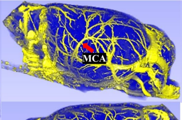
Quantitative imaging of microvasculature
Susceptibility contrast based MR structural imaging
Imaging tumor microenvironments
Funded Research Topics
Development of MR biomarkers for microvasculture studies(MISP), Imaging tumor using nano contrast agent (MISP, SRC), Multiscale imaging (UNIST)
- [1] Lee DK, Gong YL,Tessema A,Han SH, and Cho H "Resolution dependence of vessel size index across various brain regions"
NeuroImage DOI:10.1016/j.neuroimage.2024.120979 (2024) - [2] Jin S and Cho H "Cerebral hemodynamics as biomarkers for neuropathic pain in rats: A longitudinal study using a spinal nerve ligation model"
Pain DOI:10.1097/j.pain.0000000000003332 (2024) - [3] Lee H, Cho H, Lee M,Kim TH, and Lee JH "Differential effect of iron and myelin on susceptibility MRI in the subdivisions of substantia nigra"
Radiology DOI:10.1148/radiol.2021210116 (2021) - [4]
Lee DK, Kang MS, and Cho H
"MRI size assessment of cerebral microvasculature using diffusion-time-dependent stimulated-echo acquisition: A feasibility study in rodent."
NeuroImage DOI:10.1016/j.neuroimage.2020.116784 (2020) - [5] Lee H, Baek SY, Kim EJ, Huh GY, Lee JH, and Cho H "MRI T2 and T2* relaxometry to visualize neuromelanin in the dorsal substantia nigra pars compacta"
NeuroImage DOI:10.1016/j.neuroimage.2020.116625 (2020) - [6]
Lee HS, Baek SY, Chun SY, Lee, JH, and Cho H*
"Specific visualization of neuromelanin-iron complex and ferric iron in post-mortem human substantia niagra with MR relaxometry at 7T"
NeuroImage DOI:10.1016/j.neuroimage.2017.11.035 (2017) - [7] Han SH, Cho JH, Jung HS, Suh JY, Kim JK, Kim YR, Cho GG and Cho H* "Robust MR assessment of cerebral blood volume and mean vessel diameter using SPION-enhanced ultrashort echo acquisition"
NeuroImage DOI:10.1016/j.neuroimage.2015.03.042 (2015)
